Pulse site, tools and resources for University of Utah Health. Available to active employees, active students, and active POI. Pulse, rhythmic dilation of an artery generated by the opening and closing of the aortic valve in the heart. A pulse can be felt by applying firm fingertip pressure to the skin at sites where the arteries travel near the skin’s surface; it is more evident when surrounding muscles are relaxed. Different Types of Abnormal Pulses A pulse represents the arterial palpation of the heartbeat by placing fingertips at the places where an artery could be felt by pressing it against the near-surface. The radial pulse is commonly measured.
A pulse represents the arterial palpation of the heartbeat by placing fingertips at the places where an artery could be felt by pressing it against the near-surface.
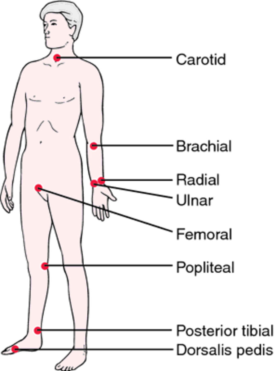
The radial pulse is commonly measured. Other sites are
- Neck – carotid artery
- Wrist- radial artery
- Groin – Femoral artery
- Knee – popliteal artery
- Ankle – posterior tibial artery
- Foot – dorsalis pedis artery
The pulse was first described by Claudius Galen.

Characteristics of Normal pulse
A pulse is generated because of the pressure waves caused by the pumping action of the heart.
It is the indirect measure of heartbeat and activity of the heart. The normal pulse has a small anacrotic wave on the upstroke which is not felt. This is followed by a big tidal or percussion wave which is felt by the palpating finger.
On the following downstroke, there is a notch followed by a wave both of which are not normally palpable.

Following are the characteristics that are looked for when palpating-
Rate
The rate is measured as beats per minute and is calculated by counting the beats for full one minute or counting for half a minute and then multiplying by two. If the rhythm is irregular, the pulse should be counted for a full one minute.
The rate varies in resting state and activity as the physiological demands vary.
Mostly, the pulse rate and heart rate are equal but in case of premature beats or atrial fibrillations, the heart rate may be more than the pulse rate
The difference is called the pulse-rate deficit.
In adults, the normal pulse appears at regular intervals and has a rate between 60-100 per min. There may be a mild variation in the rate between the two phases of respiration which is called sinus arrhythmia.
Rhythm
The normal rhythm is regular which indicates that the interval between two beats is always equal.
An irregular rhythm might indicate sinus arrhythmia.
Close attention may also indicate if the rhythm is regularly irregular [the abnormal rhythm comes at the irregular intervals] or irregularly irregular (there is no rhythm to the irregularity).
Irregularly irregular rhythm is highly specific to atrial fibrillation.
[Other causes of irregularity – ectopic beats, atrial fibrillation, paroxysmal atrial tachycardia, atrial flutter, partial heart block, etc.]
Force
Pulse force or volume is the force or strength of the pulse felt when palpating.
The force provides an idea of how hard the heart has to work to pump blood out of the heart and through the circulatory system.
The force is recorded using a scale
- 3+ Full, bounding
- 2+ Normal/strong
- 1+ Weak, diminished, thready
- 0 Absent/non-palpable

A 1+ force may reflect a decreased stroke volume [ can be seen in heart failure, heat exhaustion, or hemorrhagic shock, etc.]
A 3+ force may reflect an increased stroke volume and is seen with exercise, stress, fluid overload, and high blood pressure.
Pulse Equality
A comparison with the opposite side would give a quick idea whether the force is comparable. This value may be unequal in arterial obstructions and aortic coarctation, aortic dissection, and vascular trauma.
In coarctation of aorta or supravalvular aortic stenosis, femoral pulse may be significantly delayed as compared to radial. This is an important finding and aids in diagnosis.
Different Types of Abnormal Pulses
The normal pattern may become abnormal in different conditions. Abnormal pulses indicate a variation in heart activity. Here is a list of different types of pulses in the body.
Anacrotic
Anacrotic pulse is a slow rising, twice beating pulse where both the waves are felt during systole. The waves that are felt are the anacrotic wave and the tidal wave. It is best felt in the carotid artery in aortic stenosis.
Pulsus Bisferiens
Pulsus bisferiens is a rapidly rising, twice beating pulse where both the waves are felt during systole. Here the percussion wave is felt first followed by a small wave. It is seen in:
Idiopathic hypertrophic subaortic stenosis
In this condition, initially, there is no obstruction to the outflow and about 80 percent of the stroke volume is ejected in the early part of systole. The obstruction occurs in mid systole when aortic valve approximates the hypertrophied septum. Hence, there is a dip, as suddenly the flow ceases, followed by a secondary rise as the L.V. overcomes the obstruction.
Severe Aortic Insufficiency with mild Aortic Stenosis
The volume flow is initially increased due to severe aortic insufficiency with mild aortic stenosis. This causes an extra high-velocity jet to be shot out resulting in the second wave.
Pulsus Parvus ET Tardus
Pulsus Parvus ET Tardus is a slow rising pulse like the anacrotic pulse but the anacrotic wave is not felt. It is seen in aortic stenosis.
Pulsus Alternans

Pulsus alternans is characterized by a strong and weak beat occurring alternately, probably due to alternate rather than regular contraction of the muscle fibers of the left ventricle.
Causes are left ventricular failure, toxic myocarditis, paroxysmal tachycardias. It may occur for several beats following a premature beat.
Pulsus Paradoxus
Systolic blood pressure normally falls by 3-10 mm Hg during inspiration. This is because though there is increased venous return to the right side of the heart, there is relative pooling of the blood in the pulmonary vasculature as a result of lung expansion and more negative intrathoracic pressure during inspiration.
This decreases the venous return to the left atrium and ventricle and subsequently causes a fall in left ventricular output thereby decreasing the arterial pressure. When the drop is more than 10 mmHg, it is referred to as pulsus paradoxus. During inspiration, the pulse is erroneously called pulsus paradoxus although it is merely an exaggeration and not a reversal of the normal.
The paradox of this phenomenon is that in extreme cases the peripheral pulse can disappear on inspiration while paradoxically, heart sounds remain audible during the “missed beats”.
A reverse pulsus paradoxus may occur in patients receiving continuous airway pressure on a mechanical ventilator.
Pulsus paradoxus is seen in superior vena cava obstruction, lung conditions like asthma, emphysema or airway obstruction, cardiac conditions like pericardial effusion, constrictive pericarditis and severe congestive cardiac failure.
Pulsus Bigeminus
Pulsus bigeminus is the coupling of the waves in a pair, followed by a pause. It is seen in alternate premature beats, A.V. block, and sinoatrial block with ventricular escape.
Thready Pulse
The pulse rate is rapid and the pulse wave is small and disappears quickly. This is seen in shock especially cardiogenic.
Waterhammer Pulse
Waterhammer pulse is a large bounding pulse associated with an increased stroke volume of the left ventricle and a decrease in the peripheral resistance, leading to wide pulse pressure. The pulse strikes the palpating finger with a rapid, forceful jerk and quickly disappears. It is best felt in the radial artery with the patient’s arm elevated. It is caused by the artery suddenly emptying because some of the blood flows back from the aorta into the ventricle.
It may be seen in fever, alcohol consumption and pregnancy. It is also seen in high output states like anemia, beriberi or cor pulmonale, cirrhosis, Paget’s disease, AV fistula, thyrotoxicosis, etc.
Cardiac lesions like aortic regurgitation, rupture of sinus of Valsalva into the heart chambers, patent ductus arteriosus, aortopulmonary window, and systolic hypertension may show Waterhammer pulse as well.
1 1 - 2
1 1
An impulse can be felt over an artery that lies near the surface of the skin. The impulse results from alternate expansion and contraction of the arterial wall because of the beating of the heart. When the heart pushes blood into the aorta, the blood’s impact on the elastic walls creates a pressure wave that continues along the arteries. This impact is the pulse. All arteries have a pulse, but it is most easily felt at points where the vessel approaches the surface of the body.
The pulse is readily distinguished at the following locations: (1) at the point in the wrist where the radial artery approaches the surface; (2) at the side of the lower jaw where the external maxillary (facial) artery crosses it; (3) at the temple above and to the outer side of the eye, where the temporal artery is near the surface; (4) on the side of the neck, from the carotid artery; (5) on the inner side of the biceps, from the brachial artery; (6) in the groin, from the femoral artery; (7) behind the knee, from the popliteal artery; (8) on the upper side of the foot, from the dorsalis pedis artery.
Types Of Pulse Site
The radial artery is most commonly used to check the pulse. Several fingers are placed on the artery close to the wrist joint. More than one fingertip is preferable because of the large, sensitive surface available to feel the pulse wave. While the pulse is being checked, certain data are recorded, including the number and regularity of beats per minute, the force and strength of the beat, and the tension offered by the artery to the finger. Normally, the interval between beats is of equal length.
